
Tuesday, July 31, 2018

IBA launches SMARTSCAN beam commissioning system


SMARTSCAN
IBA's SMARTSCAN beam commissioning system on show at the AAPM Annual Meeting.
Commissioning a new linear accelerator is an intensive process, requiring a thousand or so beam measurements on a water phantom. This can take days or even weeks to complete, and the repetitive nature of this task makes it prone to user error.
To ease this procedure, IBA Dosimetry launched its SMARTSCAN beam commissioning system. Announced at this week’s AAPM Annual Meeting, SMARTSCAN guides the user through the entire linac commissioning workflow – from system setup to scanning of the entire data set – and automates repetitive tasks. As a result, beam data commissioning and accelerator quality assurance (QA) is consistently executed with optimal quality. SMARTSCAN is connected to myQA, IBA Dosimetry’s Integrated QA platform
“IBA is launching a water phantom that automates the commissioning steps,” says IBA’s Ralf Schira. He notes that there are some crucial steps where the physicist still needs control, and that SMARTSCAN provides guidance through those procedures. “It’s not a black box, it’s more like a navigation system.”
Modus - a novel method for quantifying 3D geometric distortion in MRIAdvertisementSchira explains that the development was motivated by IBA’s discussions with physicists as to what they find to be the most problematic part of beam commissioning. As well as the process taking too long and being too user intensive, many interviewees pointed out that they were not 100% confident that all of the scans were 100% correct.
“SMARTSCAN addresses both problems, by making the process more efficient and providing the required beam quality data,” says Schira. “This gives the peace of mind that beam commissioning has been done perfectly.” This, in turn, provides the foundation for safe and accurate treatment of every patient.
To ensure optimal beam data quality, SMARTSCAN checks every single scan during the process, with suspicious measurements flagged immediately. SMARTSCAN also enables commissioning work to be completed efficiently in less time, allowing faster clinical implementation of new equipment. It does this by creating the most optimal scan sequence, as well as by adapting the speed of detector motion.
“SMARTSCAN provides a great deal of intelligence and guidance,” Schira tells Physics World.

Monte Carlo accuracy

The AAPM meeting also saw IBA launch SciMoCa, a Monte Carlo-powered secondary dose check and plan verification software. Monte Carlo is generally accepted as the gold standard for dose calculation accuracy in treatment planning. Now, SciMoCa makes Monte Carlo accuracy available for secondary independent dose calculation and verification, allowing users to verify their treatment plans with an equally robust QA system.
“Introducing Monte Carlo plan QA seamlessly for all major linacs, TPS systems and treatment modalities introduces an additional level of QA accuracy in our industry,” says Jean-Marc Bothy, president of IBA Dosimetry GmbH. “At this year’s AAPM in Nashville we received exceptionally positive feedback from the medical physics community about SciMoCa’s unprecedented Monte Carlo workflow automation, calculation speed and proven accuracy”.
The FDA-cleared SciMoCa software is developed by Radialogica and Scientific RT. IBA has entered into a global distribution agreement with Radialogica.

Tuesday, July 24, 2018

Organic ferroelectrics finally stick in the memory


Xiong Ren-Gen and Yu-Meng You with their students. Credit: Yu-Meng You and Xiong Ren-Gen

Inorganic ferroelectrics have promised to change the face of semiconductor electronics for almost a century, but high processing costs have so far limited development. Now, researchers at Southeast University in Nanjing, China, have paved the way for progress by fabricating the first metal-free perovskite crystals. They present a set of materials that can achieve the performance of inorganic ferroelectrics but with the versatility, low-cost and low-toxicity inherent in organics.
To induce the directional switching of polarization characteristic of ferroelectricity, a material must contain a spontaneous dipole that can respond to an electric field. In other words, the centres of positive and negative charge within a crystal must be different. For metal-free perovskites, this should theoretically happen when a highly symmetric non-ferroelectric state is ‘frozen’ into a state with polar symmetry.


From database to device

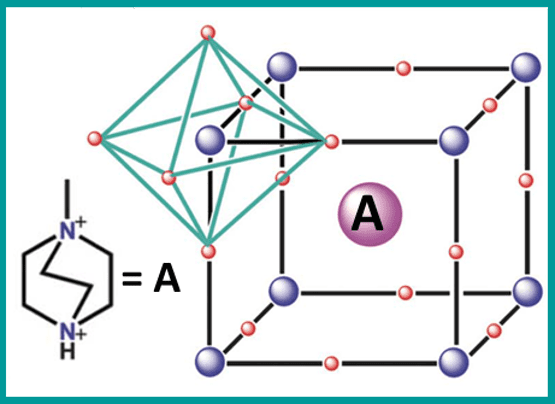
With this in mind, Ren-Gen Xiong and Yu-Meng You instructed their students to scour the hundreds of thousands of entries in the Cambridge Structural Database for molecules of suitable size and symmetry. Such candidates could then be incorporated into the traditionally metallic “A” site of the perovskite structure, yielding an all-organic perovskite ferroelectric.
The result of their efforts is the discovery of 23 metal-free perovskites including MDABCO-NH4I3(MDABCO is N-methyl-N’-diazabicyclo[2.2.2]octonium). This particular crystal displayed a spontaneous polarization of 22 microcoulombs per centimetre square, close to that of the state-of-the-art perovskite ferroelectric, BaTiO3 (BTO). In addition, crystals can be formed readily at room temperature, avoiding the excessive heat (>1000 oC) required to make inorganic ferroelectrics. This will lower fabrication costs and open the door for more delicate device applications such as flexible devices, soft robotics and biomedical devices.
The MDABCO molecule is crucial to the large spontaneous polarization that the researchers observed. At high temperatures, excessive thermal energy leaves the MDABCO molecule in a state of free rotation within the crystal. Here, the average centres of positive and negative charge at the molecule site are the same and ferroelectricity is forbidden. However, when cooled below the phase transition temperature of 448 K, the MDABCO molecule becomes locked in place revealing a significant dipole with eight possible polarization directions.

Beyond binary

Ferroelectric random access memory (Fe-RAM) works on the principle that individual cells are charged to states “0” and “1”, represented by different polarization directions of the active material.  As ferroelectric crystals tend to have two polarization states, we obtain the well-known binary system. The eight possible polarization directions in MDABCO-NH4I3 then, will pique the interest of those looking to make next-generation memory devices.
“In principle, eight polarization directions could be used to make an octonary device with eight different logic states”, explains Yu-Meng You. “This is a potential strategy for increasing the density of future RAM devices”. While You expresses concern over increased architectural complexity in such a device, the potential for cramming eight bits into a single cell could add to the commercial prospects of this set of materials.

Prospects for perovskites

But the opportunities for advancement don’t stop at memory applications. “We have demonstrated a new system of perovskites with compositional flexibility, adjustable functionalization and low toxicity. We expect the metal-free perovskite system will attract great attention in near future”.
Full details are reported in Science.

Friday, July 20, 2018

AUDITORY SYSTEM DISORDERS


Learn more about Tympanosclerosis

AUDITORY SYSTEM DISORDERS

Thomas O. Willcox, Gregory J. Artz, in Neurology and Clinical Neuroscience, 2007

Tympanic Membrane

Pathology of the tympanic membrane includes perforations, atelectasis, and tympanosclerosis. Aside from its role in protecting the middle ear, the tympanic membrane is critical in receiving sound waves and efficiently transmitting them through the ossicular chain to the endolymph of the cochlea. Any pathological process that compromises the mobility or efficiency of the tympanic membrane results in a conductive hearing loss. Tympanic membrane perforations can be caused by acute and chronic infections, head trauma, or iatrogenic causes, such as after tympanostomy tube extrusion. Tympanostomy tube placement is common in infants with otitis media and is one of the most common surgical procedures performed today. The reported rate of tympanic membrane perforation depends on the type of tube placed; however, routine grommet-type tubes have a 1% to 3% incidence.3 Most perforations from tympanostomy tubes are small, causing a 10-dB hearing loss or less, and usually heal with time. However, larger perforations and total perforations of the tympanic membrane, usually seen in patients with a history of chronic otitis media, can result in a significant conductive hearing loss of 30dB or more. A thin, atrophic, atelectatic tympanic membrane can also cause a conductive hearing loss, particularly if there is associated ossicular erosion. A retracted, atelectatic tympanic membrane is caused by eustachian tube dysfunction and the resultant chronic negative middle ear pressure. Hearing loss can be further affected in these patients by chronic middle ear fluid. Initial treatments consist of tympanostomy tube placement, tympanoplasty, and medical therapy, including decongestants and nasal steroid sprays. A common finding on otoscopy during routine physical examination is tympanosclerosis, a white discoloration of the tympanic membrane. Tympanosclerosis can be due to a prior history of tympanostomy tube placement and/or an associated history of otitis media. Unless the tympanic membrane involvement is particularly severe, it is rare for tympanosclerosis to cause an appreciable conductive hearing loss.
The ratio of the tympanic membrane area to the stapes footplate area results in an 18-fold amplification of sound under normal physiological conditions.4Any disruption or fixation of the ossicular movements impairs this efficient sound transmission. Therefore, any abnormalities of the ossicular chain manifest as a conductive hearing loss. Otosclerosis is a common cause of conductive hearing loss due to stapes footplate fixation. Otosclerosis is a disease of bone limited to the otic capsule. Classically, it causes a conductive hearing loss, but it must be mentioned that otosclerosis can also affect the cochlea, causing a mixed or even a purely sensorineural hearing loss. It is inherited in an autosomal dominant fashion with incomplete penetrance and is more often seen in white populations at a histological incidence of 7% to 10%. Only approximately 10% of patients with histological evidence of otosclerosis present with clinical symptoms.5 Two thirds of patients with otosclerosis are women. The disorder is often bilateral and classically manifests in the third and fourth decades of life as a conductive hearing loss. Otosclerosis in women has always been believed to worsen during pregnancy; however, clinical data have brought into question that premise.6 Treatment is often curative with stapedotomy or stapedectomy surgery.
Middle ear and mastoid cholesteatoma is defined as an accumulation of keratin and desquamated debris from the squamous epithelial lining of the external auditory canal and lateral surface of the tympanic membrane. There are two types: congenital and acquired. Congenital cholesteatoma is an anteriorly based mass believed to be an embryological remnant. The more common type is the acquired cholesteatoma that results from otitis media. Squamous epithelium migrates into the middle ear and mastoid. Of the acquired type, cholesteatoma can occur in the setting of a tympanic membrane perforation or from chronic otitis media due to eustachian tube dysfunction causing persistent negative pressure and tympanic membrane retraction. Cholesteatoma manifests as a middle ear mass, often in close approximation to the ossicles, with or without bony erosion, and patients present with a conductive hearing loss. Other symptoms commonly include chronic otorrhea and rarely vertigo and facial nerve paresis or palsy. Treatment consists of treating any infection first and then surgical removal of the cholesteatoma with ossicular reconstruction if warranted. Ossicular reconstruction is sometimes delayed 6 to 12 months, during which time patients are observed for any signs of recurrence or recidivism. If the posterior external auditory canal wall is left intact, recurrence rates are slightly higher at 5% to 27% versus 2% to 10% when the posterior canal wall is removed.7 When treating cholesteatoma, the first priority is to create a dry, safe ear, as infectious complications of a cholesteatoma can have significant morbidity, such as meningitis and brain abscesses. Correcting the conductive hearing loss is a second priority only after antimicrobial and surgical treatments have been successful.

Guinea Pigs as Experimental Models

Douglas K. Taylor, Vanessa K. Lee, in The Laboratory Rabbit, Guinea Pig, Hamster, and Other Rodents, 2012

Streptococcus pneumoniae

Streptococcus pneumoniae can cause a variety of clinical diseases in humans including pneumonia, bacteremia, otitis media, meningitis, sinusitis, peritonitis, and arthritis. Sequelae can include tympanosclerosis from otitis media and sensorineural hearing loss from pneumococcal meningitis (Ozcan et al., 2008; Skinner et al., 2004). These diseases can be fatal, and high-risk groups are particularly susceptible to serious disease including the elderly, children under 2 years old and those with underlying medical conditions. The use of vaccines has decreased rates of infection since 2001, and the CDC currently recommends vaccination of infants, young children, and adults (CDC, 2008c). As with other bacteria, resistant bacteria are an emerging problem and beta-lactam resistance is common. S. pneumoniae is a pathogen for many species, and has caused outbreaks in guinea pig laboratory colonies (Saito et al., 1983). This bacterium can also be found in the respiratory tract of normal animals and disease may not occur unless precipitated by a stressor. Guinea pigs are used as models of this disease primarily for research related to bacterial pathogenesis and the development of treatments. Although this generally involves the CNS or inner ear, guinea pigs have also been used to test antibiotics after experimentally induced fatal pneumonia from intratracheal instillation of S. pneumoniae (Ponte et al., 1996).
A guinea pig model of intrathecal administration has been used to evaluate pathogenesis and antibiotic effectiveness for meningitis. This model can involve intrathecal injection of bacteria into the cistern magna (Blank et al., 1994; Force et al., 2008, 2009). Others have injected bacteria into the subarachnoid space after drilling a hole over the parietal skull bone (Winter et al., 1996, 1997). Infection can lead to labyrinthitis, meningitis, and death. After infection, cerebrospinal fluid (CSF) is taken at various time points to assess for inflammation, protein, and bacterial counts. Blood may be evaluated to assess for bacteremia. Other parameters measured may include auditory integrity and evaluation of the organ of Corti by electron microscopy or histology (Blank et al., 1994; Winter et al., 1996, 1997). This model has been used to test antibiotics such as meropenam, rifampin, ceftriaxone, and vancomycin (Force et al., 2008, 2009; Nairn et al., 1995). Force et al. (2008)compared the guinea pig to the traditional rabbit model of meningitis. The disadvantage of the guinea pig is that their small size only allows for CSF collection at a maximum of two time points, whereas rabbits allow collection of a larger volume. However, the authors suggest that guinea pigs are a more reliable model for testing efficacy of certain drugs. For example, rabbits easily hydrolyze the drug meropenam and metabolize it more quickly than guinea pigs and humans.
Guinea pigs have also been used to study the otoxicity of S. pneumoniae by direct administration into the ear as a model of otitis media. Bacteria or pneumococcal proteins are inoculated through the tympanic cavity and directly into the middle ear or cochlea. Parameters often measured include otomicroscopic exams and auditory nerve function, followed by euthanasia for histology and bacterial cultures (Cook et al., 1999; Hori et al., 2000; Ozcan et al., 2008; Skinner et al., 2004; Winter et al., 1998). This model has also been used to evaluate treatments such as topical doxycycline (Hori et al., 2000; Ozcan et al., 2008; Winter et al., 1998).

Ear and Temporal Bone

BRUCE M. WENIG, in Modern Surgical Pathology (Second Edition), 2009

Otitis Media

Otitis media is either an acute or chronic infectious disease of the middle ear space. Otitis media is predominantly but not exclusively a childhood disease. The most common organisms implicated in causing disease are Streptococcus pneumoniaeand Haemophilus influenzae. Otoscopic examination reveals a hyperemic, opaque, bulging tympanic membrane with limited mobility; purulent otorrhea may be present. Bilateral involvement is not uncommon. The middle ear infection is thought to result from infection via the eustachian tube at the time of or following pharyngitis (bacterial or viral). In general, otitis media is managed medically. However, tissue is sometimes removed for histopathologic examination. The pathologic alterations are generally straightforward, but secondary changes such as glandular metaplasia of the surface epithelium, the result of chronic infection, may occur, causing confusion with a true gland-forming neoplasm.
There are no specific macroscopic features. Tissue specimens are usually received as multiple small fragments of soft to rubbery granulation-type tissue. If tympanosclerosis is present, the tissue may be firm to hard, consisting of calcific debris. In general, all tissue fragments should be processed for histologic examination. The histology of otitis media varies and depends on the disease state.148 Acute otitis media is virtually never a surgical disease. The inflammatory infiltrate in acute otitis media is composed of polymorphonuclear leukocytes. Acute inflammatory cells may be superimposed in chronic otitis media. The histologic changes in the chronic form include a variable amount of chronic inflammatory cells consisting of lymphocytes, histiocytes, plasma cells, and eosinophils. Multinucleated giant cells and foamy histiocytes may be present. The middle ear low cuboidal epithelium may or may not be seen. However, glandular metaplasia (Fig. 13-13), a response of the middle ear epithelium to the infectious process, may be present. These glands tend to be more common in nonsuppurative than in suppurative otitis media. The metaplastic glands are unevenly distributed in the tissue specimens, variably shaped, and separated by abundant stromal tissue. The glands are lined by a columnar to cuboidal epithelium, with or without cilia or goblet cell metaplasia. Glandular secretions may or may not be present; thus the glands may appear empty or may contain various secretions, including thin (serous) or thick (mucoid) fluid. The identification of cilia is confirmatory of middle ear glandular metaplasia and is a feature not found in association with MEA.149 In addition to the inflammatory cell infiltrate and glandular metaplasia, other histopathologic findings usually seen in chronic otitis media include fibrosis, granulation tissue, tympanosclerosis, cholesterol granulomas, and reactive bone formation.
Tympanosclerosis represents dystrophic mineralization (calcification or ossification) of the tympanic membrane or middle ear and is associated with recurrent episodes of otitis media.150Tympanosclerotic foci may be localized or diffuse and appear as white nodules or plaques. Histologically, dense “clumps” of mineralized, calcified, or ossified material or debris can be seen within the stromal tissues or in the middle (connective tissue) aspect of the tympanic membrane. Tympanosclerosismay cause scarring and ossicular fixation.
Cholesterol granulomas represent a foreign body granulomatous response to cholesterol crystals derived from the rupture of red blood cells that accompaines breakdown of the lipid layer of the erythrocyte cell membrane. Cholesterol granulomas arise in the middle ear in any condition in which there is hemorrhage combined with inadequate drainage and ventilation of the middle ear space.151 The histology of cholesterol granulomas includes the presence of irregular-shaped, clear-appearing spaces surrounded by histiocytes or multinulceated giant cells (foreign body granuloma). Cholesterol granulomas are not related to cholesteatomas but may occur in association with or independent of them. Tympanosclerosis and cholesterol granulomas may occur independent of otitis media; cholesteatomas may or may not be associated with otitis media.
The differential diagnosis of the glandular metaplasia seen in otitis media includes MEA. The haphazard arrangement of the glands and the presence of cilia against the background of changes of chronic otitis media should allow the differentiation of metaplastic from neoplastic glands.
In the antibiotic era, complications associated with otitis media are rare. However, if left unchecked, complications of otitis media include acute mastoiditis, suppurative labyrinthitis (inflammation of the inner ear), meningitis, and brain abscess.

Urology

Jerilyn M. Latini MD, ... William W. Roberts MD, in Clinical Men's Health, 2008

Peyronie's Disease

Definition

Peyronie's disease is an acquired inflammatory condition of the penis resulting in penile curvature. Additionally, it can be associated with penile pain or erectile dysfunction. In general, the disease progresses through two phases, which include an initial phase consisting of pain and gradual penile deformity followed by resolution of the pain with stabilization of the penile plaque and deformity. It is estimated that the incidence of symptomatic Peyronie's disease is approximately 1%.56 In white men, the average age of onset is 53 years.

Etiology and Risk Factors

It is generally believed that Peyronie's disease results from trauma that results in abnormal stimulation of the wound-healing process and subsequent scar formation in the tunica albuginea. Several disease entities are also associated with Peyronie's disease, including Dupuytren's contractures, plantar fascial contractures, tympanosclerosis, diabetes mellitus, gout, and Paget's disease, as well as use of phenytoin and beta-blockers.57

Evaluation

For men presenting with Peyronie's disease, the history should include disease duration, degree of curvature, whether penetration with intercourse is still possible, and whether concomitant pain or erectile dysfunction is present. Additionally, one should inquire about any history of trauma or other risk factors present above. The physical examination should focus on evaluation of the patient's hands and feet for contractures as well as palpation of the penile plaque. Typically, the plaque will be located on the dorsum of the penis, although plaques can also be lateral or ventral.

Treatment

There are few RCTs that have examined therapies for Peyronie's disease. In general, medical therapy is the first-line treatment for men with newly diagnosed disease. Surgical intervention is reserved for men with a stable plaque/penile deformity for at least 6 months who are unable to engage in intercourse or have concomitant erectile dysfunction unresponsive to nonsurgical therapies.
Multiple medical therapies have been used to treat Peyronie's disease. Vitamin E, given as 800 IU/day, may be effective. It is commonly given because of its antioxidant properties and general tolerability, but no RCT has been performed to demonstrate efficacy over placebo. Potassium aminobenzoate (Potaba), given at 12 g/day (divided in 4–6 doses), has been demonstrated to be efficacious in a small blinded study58; however, gastrointestinal upset and the large quantity of oral pills limits its widespread use. Other agents that have been used include colchicine, but results are only anecdotal. A small RCT evaluated tamoxifen versus placebo and found no difference in symptomatic outcomes.59 Several investigators have reported successful treatment of Peyronie's disease by injecting calcium channel blockers,60 steroids,61 and interferons62 into the penile plaques, although no RCTs on the topic have been performed to date. A small RCT comparing intralesional collagenase and placebo demonstrated improvement in mild to moderate Peyronie's plaques, although there was not a statistically significant improvement for more severe curvature.63
Several surgical options exist to correct the penile curvature from Peyronie's disease, although no RCTs have been performed to compare these treatments. Plication of the tunica albuginea on the contralateral side of the corpora cavernosa bend is effective in straightening the penis; however, men will lose some penile length from this procedure. To avoid this, an alternative option is incision or excision of the plaque with grafting using either autologous or synthetic materials. The drawback to this approach is that oftentimes the neurovascular bundle on the dorsum of the penis must be mobilized, which can cause penile numbness and may be permanent. Finally, for men with concomitant erectile dysfunction, a penile prosthesis can be placed and the penis straightened with intraoperative molding.64

Tympanoplasty—Staging and Use of Plastic

James L. Sheehy, Clough Shelton, in Otologic Surgery (Third Edition), 2010

INDICATIONS FOR STAGING

There are two reasons for staging the operation in tympanoplasty: (1) obtaining a permanently disease-free ear and (2) obtaining permanent restoration of hearing.3,4 Whether one finds any indication for staging depends on how vigorously a good functional result is pursued in badly diseased ears.
The decision whether or not to stage is made at the time of surgery. With experience, one usually can make this judgment preoperatively and alert the patient to the possible necessity of a two-stage procedure. The decision is based on three factors: (1) the extent of the mucous membrane problem, (2) the certainty (or lack thereof) of removal of cholesteatoma, and (3) the status of the ossicular chain. Taking these three factors into account, we stage about 75% of tympanoplasty and mastoidectomy procedures and about 15% of tympanoplasties not requiring mastoidectomy.

Mucosal Disease Factors

There are frequently large areas of diseased or absent mucosa in the chronically infected middle ear. Groundwork is necessary to promote regrowth of normal mucosa. The first step is elimination of infection before surgery, if possible. The second step is removal of all squamous epithelium, granulations, and irreversibly diseased mucosa at the time of surgery. The middle ear is sealed with a graft to prevent squamous epithelium from migrating back into the middle ear. This sealed middle ear space fills with a blood clot, and this clot supports fibroblastic invasion with eventual formation of scar tissue or adhesions between the denuded surfaces. To prevent these adhesions from forming, and to allow mucosa to migrate in, plastic sheeting is used over the denuded areas.
A two-stage operation is indicated to obtain the best hearing results, and to prevent recurrence of cholesteatoma (retraction pocket) in patients with extensive mucous membrane destruction. The objective of the two-stage procedure is to obtain a well-healed ear with a mucosa-lined pneumatized middle ear cleft so that ossicular reconstruction may be performed later under ideal circumstances.

Ossicular Chain Factors

An increase in the incidence of sensorineural hearing impairment has been observed in patients in whom the inner ear has been opened in the presence of actual or potential infection. Because of this result, a fixed stapes should not be removed at the time of tympanic membrane grafting. In cases of otosclerosis, a two-stage procedure is almost always indicated. When the fixation is due to tympanosclerosis, it may be possible to mobilize the stapes, depending on the area of fixation. In diffusely involved cases, a laser can be useful to remove the suprastructure and to char the tympanosclerosis, allowing its removal and resulting in a mobile footplate. If it is impossible to mobilize the footplate, a second stage procedure should be carried out. At that time, the tympanic membrane would be intact, and the middle ear would be without infection, allowing for removal of the fixed footplate in a sterile environment and completion of ossicular reconstruction.

Residual Cholesteatoma Factor

It may seem illogical to leave behind epithelial disease, removing it at a planned second stage procedure, but this is exactly what is done under certain circumstances. Removal of cholesteatoma in the middle ear may be questionable sometimes in an acutely inflamed ear, in which differentiating between granulation tissue and matrix is difficult. Differentiating becomes a particular problem when granulations fill the oval and round windows. Excessive manipulation in these areas could result in an inner ear complication.
Removal of matrix involving a mobile stapes with an intact suprastructure can be challenging. Sometimes it is impossible to be certain that every shred of cholesteatoma has been removed. In such cases, a laser can be used at the second stage to cut the crura off the mobile footplate and facilitate removal of residual disease.
The surgeon may have torn the matrix when removing it from the tympanic recess and may be uncertain of complete removal, which presents a considerable problem under the pyramidal process, an area hidden from view regardless of the technique of surgery, whether it is an open or closed cavity technique. Removal of the pyramidal process with a diamond burr may or may not resolve the problem. One third of patients with middle ear cholesteatoma at the first operation have residual disease at the second stage.2
It is much easier to be certain of cholesteatoma removal from the mastoid, especially in a small apneumatic one. Extensive cholesteatoma in a pneumatized mastoid poses a problem. In using the intact canal wall procedure, one should usually revise the mastoid in such cases within 1 to 2 years to be certain not to leave disease behind.
The mastoid and epitympanum are often re-explored in patients in whom excessive bleeding occurred at surgery. Unexpected residual disease in the epitympanum has been noted in some cases of this type in the past.

Timing the Second Stage

The second-stage operation may be performed in 6 to 9 months if the primary indication for staging was an ossicular or a mucous membrane problem. The middle ear should be well healed by that time. If the primary reason for staging is reinspection of the mastoid and epitympanum for possible residual cholesteatoma, it is best to wait 9 to 18 months. The delay allows time for any residual disease to have grown to a 1 or 2 mm cyst so that it may be identified with greater ease. The only exception to this rule is if this disorder occurs in a child, or if serous otitis media develops; a residuum may grow faster under these circumstances.
What is the best strategy for managing a patient with bilateral cholesteatomas who needs staged procedures on both ears? The decision here is based on disease activity and hearing level. After the first stage, it is common for the ear to have a maximal conductive hearing loss until the second operation. This ear would not provide the patient with functionally useful hearing without a hearing aid.
If only one ear has active disease or poor hearing, that ear is operated initially. After the second stage is completed, surgery begins on the second ear. If both ears have active disease, after healing has occurred from the first stage on the initial ear, the second ear can be operated. The patient typically requires a hearing aid on the first ear until the ossicular reconstruction is performed at the second stage. Fitting a behind-the-ear hearing aid provides the capability to switch the aid easily to the opposite ear as needed.
Patients with chronic otitis media present the surgeon with many management complexities. Bilateral disease is common, and each ear may require two surgeries. It is helpful to record the plan for the second stage at the time the first stage is completed; this can be done through a handwritten chart note, a customized surgical data sheet, or prominent placement in the dictated operative report. Common included items are timing of the next stage, approach (transcanal or postauricular), type of prosthesis, location of possible residual cholesteatoma, need for special equipment (laser, facial nerve monitor), and areas of special caution (exposed dura, dehiscent facial nerve or jugular bulb). These details are best documented at the time of the first operation, and provide a clear basis for the planning of the second stage.

EXAMINATION OF HEARING AND BALANCE

Brian C. Kung, Thomas O. Willcox, in Neurology and Clinical Neuroscience, 2007

Physical Examination

A complete head and neck examination can give many clues to the cause of a patient's hearing loss. The auricle and the postauricular area should be examined for deformities, surgical incisions, the presence of a hearing aid, and patency of the external auditory canal. Something as simple as cerumen impaction can be the cause of hearing loss in some patients, but other conditions, such as foreign bodies, exostoses, canal stenosis/atresia, and carcinoma of the external canal, can be more troublesome. Pneumatic otoscopy can then be used to examine the tympanic membrane and middle ear. Here, the presence of a tympanostomy tube, tympanosclerosis (scarring of the tympanic membrane), tympanic membrane perforation, retraction pocket, fluid in the middle ear, middle ear masses, or otorrhea can be assessed. It is important to obtain a good seal with the speculum in order to assess the mobility of the tympanic membrane. External and middle ear abnormalities usually point to a conductive component of hearing loss.
Tuning fork testing is an essential part of the physical examination and can help determine if the cause of hearing loss is conductive, sensorineural, or mixed. The three types of tuning forks that can be used are 256Hz (middle C), 512Hz (octave above middle C), and 1024Hz (two octaves above middle C). The Rinne test is useful in determining if there is a conductive hearing loss and is performed by striking the tuning fork and placing it on the mastoid bone (testing bone conduction). Once the patient stops hearing the sound, the tines of the tuning fork are then placed in front of the external canal (testing air conduction) with the tines oriented in the head-frontal plane, and the patient indicates whether he or she can hear the sound. If the patient can hear the sound, air conduction is greater than bone conduction, and the result is normal, or “positive.” If the patient cannot hear the sound, bone conduction is greater than air conduction, and the result is abnormal, or “negative.” The degree of conductive hearing loss can be estimated based on the results of the Rinne test. A test that is negative at 256Hz and positive at 512 and 1024Hz indicates a mild 20- to 30-decibel (dB) conductive loss. A test that is negative at 256 and 512Hz and positive at 1024Hz indicates a moderate 30- to 45-dB conductive loss. A negative test at all three frequencies indicates a severe 45- to 60-dB conductive loss.6,7The Weber test is a test used to lateralize the hearing loss. The tuning fork is struck and placed on the patient's vertex, nasal bones, or maxillary teeth in the midline. The single most clinically useful fork used here is the 512-Hz variety, as the 256-Hz fork can be overly sensitive, leading to many false-positive results, and the 1024-Hz fork may not be sensitive enough.7–9 Lateralization of sound to one ear during the Weber test indicates either a conductive hearing loss in that ear or a greater sensorineural loss in the opposite ear.7Simple tuning fork tests using only a few frequencies are far from comprehensive. If both ears are symmetrically affected by a sensorineural hearing loss, both the Rinne and Weber tests will be normal, provided the patient is able to hear the tuning fork at all.
The physical examination should also include an assessment of any craniofacial deformities or stigmata that may be associated with hereditary causes of hearing loss or associated systemic diseases. Also, a full cranial nerve examination should be performed, as asymmetries in any of the cranial nerves may indicate that hearing loss is just one component of more severe or extensive disease, such as a skull base neoplasm. A decreased corneal blink reflex and hypesthesia of the external auditory canal (Hitselberger's sign) can be suspicious for an acoustic neuroma. Finally, attention to the nose, nasopharynx, oral cavity, oropharynx, larynx, and hypopharynx can reveal other causes of hearing loss (e.g., the presence of nasopharyngeal carcinoma as the cause of serous otitis media).

Acute otitis media

David M. Spiro MD MPH, Donald H. Arnold MD MPH, in Berman's Pediatric Decision Making (Fifth Edition), 2011

Clinical approach

A.
In the patient history, ask about earache and discharge, fever, respiratory symptoms, conjunctivitis, irritability, crying, decreased feeding, difficulty sleeping, vomiting, and ataxia. Inquire about the timing and treatment of the most recent AOM episode and frequency of episodes during the past 6 and 12 months. Ear tugging and other nonspecific symptoms are not reliable predictors of AOM. Identify children with immune disorders, acquired immunodeficiency syndrome, cystic fibrosis, or Kartagener syndrome (immotile cilia).
B.
Examine the TM with pneumatic otoscopy. Use a pneumatic otoscope head and remove sufficient cerumen to have an adequate view of the membrane. It is important to create an adequate seal with an appropriately sized speculum, to have adequate light intensity with a halogen light source. Signs of AOM are bulging contour with exudate (bulging pus), and diminished or absent eardrum mobility. On occasion, bullae form between the outer and middle layers of the TM (bullous myringitis). Look for signs of middle-ear damage, such as tympanosclerosis (chalky white deposits in the eardrum), retraction pocket, perforation, or cholesteatoma (yellow greasy mass). Examine the mastoid area behind the ear for tenderness, swelling, protrusion of the ear, or erythema.
C.
The most important step is to establish the proper diagnosis of AOM. Antibiotic therapy is not recommended for otitis media with effusion or for upper respiratory infections. The American Academy of Pediatrics developed a clinical guideline that establishes a diagnosis of AOM based on three components:
1.
Acute onset (<48 hours) of signs and symptoms,
2.
Middle-ear effusion, and
3.
Signs and symptoms of middle-ear inflammation
D.
The pain associated with AOM should be uniformly treated. Antibiotics have no analgesic properties. A small number of trials have demonstrated the short-term effectiveness of topical analgesic drops such as benzocaine-antipyrine and lignocaine. Systemic analgesics such as ibuprofen have been shown to improve symptom relief and may improve clinical outcomes. When pain is severe, tympanocentesis results in prompt relief.
E.
Immediate treatment of AOM with antibiotics is controversial because of questions about efficacy and concerns about increasing the prevalence of drug-resistant microbes. A meta-analysis of the results of 33 randomized clinical trials found that treatment with antibiotics increased the resolution rate by only 13.7%. First-line therapy in a nonallergic child is high-dose amoxicillin (Table 1). High-dose amoxicillin will usually eradicate the most invasive pathogen, S. pneumoniae, and if no improvement occurs, a second-line antibiotic may be chosen to cover M. catarrhalis and β-lactamase–producing H. influenzae. Consider an initial single intramuscular dose of ceftriaxone when the patient is unable to take oral antibiotics because of vomiting. If the patient is allergic to penicillin or amoxicillin, treat with azithromycin when there is a history of a serious type I hypersensitivity reaction (severe urticaria, respiratory distress, anaphylaxis), or with cefdinir, cefuroxime, or cefpodoxime when there is a history of a less severe reaction. Antihistamines, decongestants, and steroids are of no benefit in treatment of AOM.
F.
The optimal duration of amoxicillin therapy is controversial. According to Dagan and colleagues (2001), using a shorter duration regimen of 5 to 7 days appears to decrease drug-resistant pneumococcal carriage after therapy. This is important because nasopharyngeal flora determines the pathogen of the next AOM.
G.
Most clinicians in the United States routinely treat AOM with antibiotics. Some European countries frequently use a wait-and-see, observational approach to avoid unnecessary use of antibiotics, with similar rates of mastoiditis compared with the United States. Counsel parents of untreated children who do not improve within 48 to 72 hours of diagnosis to fill the prescription or return to be reassessed and treated with antibiotics if the AOM is immediately treated with an antibiotic. Approximately two of every three families will fill the wait-and-see prescription. Observational therapy should be considered when children do not meet high-risk criteria: ill appearance, recent AOM diagnosis, limited access to medical care, compromised immunity, concurrent antibiotic use, or an identified bacterial infection other than AOM.
H.
A scheduled re-examination of the ear is usually not necessary for the previously healthy child.
I.
Risk factors for unresponsive infections include age younger than 18 months, a history of recurrent AOM in the child or a sibling, and a history of antibiotic treatment of AOM within the preceding month. Parents of children older than 15 months usually know when their child’s infection has resolved. Consider treating unresponsive AOM with amoxicillin-clavulanate, cefdinir, cefuroxime, or cefpodoxime to cover both S. pneumoniae and β-lactamase–producing pathogens. This type of second-line agent is also indicated when a child experiences another symptomatic infection within 4 weeks of stopping amoxicillin; however, repeated use of amoxicillin is indicated if more than 4 weeks have passed without symptoms because a new pathogen is usually present.
J.
If a child remains symptomatic longer than 3 days while taking a second-line agent, consider performing a tympanocentesis or treating with oral clindamycin (effective against resistant pneumococci but not β-lactamase–producing organisms) or intramuscular ceftriaxone (one dose a day for 3 days; effective against both resistant pneumococci and β-lactamase–producing organisms). Tympanocentesis is performed by placing a needle through the TM and aspirating the middle-ear fluid. Indications for a tympanocentesis or myringotomy are: (1) AOM in an infant younger than 6 weeks with a past neonatal intensive care hospitalization, because the pathogens may be gram-negative; (2) AOM in a patient with compromised host resistance, because the organism may be unusual; (3) unresponsive AOM despite courses of two to four different antibiotics; (4) acute mastoiditis or suppurative labyrinthitis; and (5) severe pain.

Surgery of Ventilation and Mucosal Disease

Bradley W. Kesser, ... Rick A. Friedman, in Otologic Surgery (Third Edition), 2010

Technical Details

Tympanostomy Tube Insertion

The ear canal is gently cleaned of all wax and debris. Contact with the anterior bony canal wall is avoided because of risk of bleeding. The tympanic membrane is inspected, and the short process of the malleus is identified. This is a constant landmark and may be the only one available in cases of acute infection. The tympanic membrane is incised anteroinferiorly by using an incision that parallels the fibrous annulus (see Fig. 6-1). Use of a radial incision is satisfactory, but may be limited by an overhanging anterior canal wall. Posterior incisions should be avoided because they place the ossicles at risk. The incision is gently spread open. Care is taken to avoid any major vessel in the tympanic membrane to prevent hemorrhage into the layers of the eardrum. This bleeding into the drum is thought to predispose to tympanosclerosis.
The middle ear should be evacuated by using a small-diameter (5 Fr) suction cannula. Occasionally, gluey material is too viscous to pass through the cannula. We do not recommend using anything larger than the 5 Fr cannula. In these cases, the middle ear and ear canal can be irrigated with warm sterile saline. This usually breaks the viscous material up enough to pass through the cannula. Not all of the effusion needs to be evacuated; as long as the middle ear has a near-normal airspace to place the tube, the remainder of the effusion is carried into the Eustachian tube or drains out the tube. Culture of the effusion rarely is done.
It is important to position the tube such that the lumen is in line with the surgeon’s line of sight, facilitating postoperative examination of the middle ear mucosa in the office and cleaning of the tube should it become plugged later on. When using T-tubes, the surgeon should ensure that the short arms of the tube are completely unfolded. Ototopical drops are placed if there is an acute infection. A small cotton ball is placed in the meatus.

Laser Myringotomy

The laser has become a useful, albeit expensive, tool in the management of chronic OME. Advantages of the laser include office-based application, ease of use, and the ability to place a controlled perforation in the tympanic membrane that stays open for a medium length of time (2 to 6 weeks). Using the CO2 laser at 12 W with a single 100 ms pulse through a 200 mm objective, Goode73reliably placed 1.5 to 2 mm perforations in the tympanic membranes of 10 subjects. Ten of the 11 ears healed within 6 weeks. Tube placement was avoided.
Marchant and Bisschop74 performed 20 consecutive CO2 laser myringotomies on ears with chronic OME. All myringotomies closed within 4 weeks, with an average closing time of 17 days; 60% of cases of chronic OME were cured after 3 months. CO2 laser myringotomy has application in clinical situations when middle ear ventilation is needed for a medium length of time (weeks) without having to place a ventilation tube. Disadvantages include cost, need for extra machinery that can be bulky, required maintenance, instruction on use and technique, and office space. Local anesthesia (iontophoresis or topical phenol application) is still required. Most otologists prefer simple cold-steel myringotomy with or without tube placement, but CO2 myringotomy is an alternative; only time and experience will tell whether this technique will have widespread application and use.

Adenoidectomy

For an adenoidectomy, the patient is given a general anesthetic, and the airway is secured via endotracheal intubation. The patient is placed in the Rose position with the neck extended over a shoulder roll and draped, as described earlier. The mouth gag is inserted and suspended from the Mayo stand located over the body of the patient. The soft palate is retracted with red rubber catheters. The hard and soft palates are palpated for the presence of a submucous cleft palate. The adenoid pad is inspected with the curved laryngeal mirror.
The adenoid is excised with curved curettes of various sizes (Fig. 6-2A). The curette is seated high in the nasopharynx, and the adenoid pad is resected with a down-sweeping motion on the curette. Care must be taken to avoid injury to the prevertebral fascia and muscles, which may cause excessive bleeding. The nasopharynx is palpated for residual adenoid tissue; a second or third pass may be necessary. Curved biting forceps are useful to remove tissue inaccessible by the curette. The mirror is again used to inspect the site. Curettage of the tissue in the fossa of Rosenmüller is not done because it may lead to scar tissue formation and contracture that might result in stenosis or Eustachian tube reflux or both. Direct injury to the Eustachian tube also is avoided. The goal of surgery is the complete removal of the midline adenoid pad to achieve smooth re-epithelialization of the nasopharynx.
Bleeding usually stops quickly; tonsil sponges are used to pack the nasopharynx for hemostasis. The nasal cavities and nasopharynx are irrigated with warm saline. A malleable suction cautery can be used for precise coagulation, but its use is cautioned because of the risk of stenosis (see Fig. 6-2B).

Mastoidectomy

The ear canal and postauricular areas are initially injected with 1% lidocaine with 1:100,000 concentration of epinephrine. Vascular strip incisions are started medially at the fibrous annulus and carried laterally along the tympanomastoid and tympanosquamous suture lines (approximately 12 and 8 o’clock positions for a right ear and 12 and 4 o’clock positions for a left ear). The incisions should come over the bony-cartilaginous junction laterally. The incisions are connected medially around the annulus with the round knife, and the vascular strip is elevated from medial to lateral. A cotton ball soaked in 1:100,000 epinephrine solution (with or without lidocaine) is placed in the canal, and attention is turned to the postauricular area.
The postauricular incision is based about 1 fingerbreadth behind the postauricular crease, roughly paralleling the free margin of the helix (Fig. 6-3). The further posterior the incision, the greater the ease of inspecting the middle ear and Eustachian tube through the facial recess. The incision is carried slightly anterior in its inferior dimension to allow the ear to be retracted forward easily. In a child, care is taken not to extend the incision beyond the mastoid tip, which is more superior than in an adult. Carrying the incision more inferior or anterior puts the facial nerve at risk. Superiorly, the temporalis fascia is identified, and a piece of fascia is harvested if needed. The fascia identifies the plane of dissection. The ear is held forward with a self-retaining retractor. The linea temporalis is palpated, and an incision is made down to the bone along this line from anterior to posterior. A second incision is made perpendicular to the first in a curvilinear fashion down to the mastoid tip. The Lempert elevator is used to elevate the periosteum to identify the cribriform area and posterior canal wall. A small elevator is next used to elevate the vascular strip out of the canal. The vascular strip is held forward with the ear under the self-retaining retractor. The tympanic membrane is carefully elevated, and the middle ear is inspected.
A large cutting burr and continuous suction-irrigation are used to remove the lateral mastoid cortex. Important landmarks to identify include the posterior bony canal wall anteriorly, the tegmen mastoideum superiorly, the sigmoid sinus posteriorly, and the digastric ridge inferiorly. Care is taken not to expose the dura of the middle cranial fossa or the sigmoid sinus. When these lateral landmarks have been identified, the dissection is continued medially under the microscope. Körner’s septum is opened medially, and the mastoid antrum is identified. This dissection is carried anteriorly to open the aditus ad antrum and attic. The fossa incudis and short process of incus are carefully uncovered. The short process of the incus should be seen refracted through water. The short process marks the level of the facial recess. All air cells of the mastoid cortex should be taken down to reduce the surface area of the system. The bony plates over the posterior and middle fossa dura are skeletonized to form a smooth surface (Fig. 6-4). Care is taken to avoid exposing dura. The mucosa regenerates into a single large cavity.
The descending segment of the facial nerve is identified (using a diamond burr and copious suction-irrigation) by gentle dissection from superior to inferior using the fossa incudis, lateral semicircular canal, and digastric ridge as essential landmarks. The nerve and blood vessels on the nerve can be seen through bone. Care is taken not to expose the nerve.
The facial recess is opened into the middle ear by the use of progressively smaller diamond burrs. The plane of the short process of the incus leads to the facial recess (Fig. 6-5). When the fallopian canal and chorda tympani nerve are identified, the dissection is carried between them medially to open into the middle ear. Coupled with the tympanotomy, all parts of the mesotympanum can be inspected. The ossicular chain is palpated, and any hyperplastic mucosa, granulation tissue, or secretory tissue is removed. If bone of the middle ear/promontory is exposed, a piece of absorbable gelatin film (Gelfilm) is placed through the facial recess and across the promontory toward the Eustachian tube at the end of the procedure to prevent adhesions and to keep the recess open.
When all hyperplastic mucosa has been removed, the tympanic membrane is folded down back over the bony annulus and grafted if necessary. Cortisporin-soaked absorbable gelatin sponge (Gelfoam) packing is placed in the medial canal. The vascular strip is returned to the posterior canal wall, and the periosteal flap is resutured to the native periosteum with 2-0 chromic suture. The vascular strip is inspected transcanal and carefully placed back so that no edges are rolled under. The remainder of the canal is packed with Cortisporin-soaked Gelfoam. The postauricular incision is closed meticulously with 3-0 undyed (Vicryl) in the subcutaneous layer, avoiding the need for skin sutures. A small Penrose drain can be placed in the mastoid and carried out through the inferior aspect of the incision if the mastoid is very weepy. The drain is removed when the mastoid no longer drains. A cotton ball is placed in the meatus, Steri-Strips are applied to the postauricular incision, and a sterile dressing consisting of Telfa, gauze, fluffs, and a mastoid (or cup) wrap is placed.